NON-DOMESTIC SPP

How to use these images


IMPORTANT: To see an enlarged image, click on any image you see in LORI. Then, RIGHT click on the enlarged image to save it at its full size.


Saturday, November 30, 2019

A brief look at the family Leporidae (hares and rabbits)

Keywords: Leporidae, Oryctolagus, Lepus, Sylvilagus, anatomy, physiology, reproduction, uterus, ovaries, tract, vagina, rabbits, hares

A fascinating group of mammals, some familiar to veterinary and animal science, others not. Note that  rabbits and hares belong to the order Lagomorpha, of which there are two families: Leporidae and Ochotonidae (pikas, mouse hares, or conies)

As mentioned in the LORI entry on semen collection in rabbits, rabbit farming is underdeveloped in North America compared with Europe, Asia and South America. Therefore rabbits are superficially addressed in most animal science and veterinary curricula in North America. However, rabbits deserve attention as important sources of meat and fiber and their unique value as subjects for scientific study (See: Foote and Carney, 2000). 

Vocabulary:

Lagamorph: A member of the order Lagamorpha
Leporid: A member of the family Leporidae, comprising the rabbits and hares.
Kindling: The name given to parturition in rabbits and hares.
Kits: Perinatal rabbits and hares (the term "pup" is reserved for rats, mice and dogs).
Doe: Adult female Leporid
Buck: Adult male Leporid.
Nests and burrows: Hares (always wild) live in nests, above ground. Wild rabbits such as Cottontails live in burrows.

Introductory notes: Rabbits and hares are all members of the family Leporidae.  This family includes the domestic rabbits (Oryctolagus cuniculus), hares (many Lepus spp) and brush rabbits (many Sylvilagus spp). They share similar reproductive characteristics but as donkeys are to horses, they are also distinctively different. For example, hares and brush rabbits have 48 chromosome and precocious neonates with open eyes, able to ambulate efficiently. By contrast, rabbits have 44 chromosomes and altricial young, blind, hairless and unable to ambulate. Their eyes open at about 10 days postpartum.

In the course of this entry, the author will entertain other differences between hares and rabbits. 

Figure 1. The reproductive tract of a Snowshoe hare (Lepus Americanus) that was approximately 14 days pregnant. The ~20 day old fetus at left is that of a domestic rabbit (Oryctolagus cuniculus).  The 14 day pregnancy shows the bulbous embryo-placental units typical of rabbits and hares. Note too, that the ovaries are not covered by ovarian bursae. Corpora lutea of pregnancy are visible within the ovaries. Finally, note the double cervix (inset at right); typical for all Leporids (as well as rats and mice). The vagina (not shown) is about 10 to 12 cm long. Image size: 1344 x 831 px

A crash course on rabbit and hare reproduction: 
1. Puberty occurs between 4 and 7 months of age; later in large rabbit breeds such as the Flemish White. 

2. Although commercial rabbits breed throughout the year, hares and wild rabbits (Sylvilagus spp; Cottontailsare like cats. i.e. they respond to increasing day length and breed mostly during spring and early summer. 

3. Like cats, rabbits and hares do not have discrete estrous cycles. Instead, as reflected by vaginal cytology, follicle waves mature every 4 to 5 days (~ 7 days in hares). 

4. Although spontaneous ovulation can occur, ovulation is generally induced by copulation (mounting in the absence of intromission is also thought to be important). Ovulation occurs about 10 hours post copulation. GnRH treatment is essential for satisfactory pregnancy rates when AI is used.

5. Pregnancy diagnosis is accurate at 10 to 14 days post-breeding using transabdominal palpation. Optimal first service conception rates should be greater than 70%  i.e. 70% of does should be pregnant after a single breeding.

6. Gestation is longer in hares than rabbits; about 42 days vs. 30 days. This explains why hares have precocious young and rabbits, altricial young. 

7. Parturition occurs in the morning. Anterior, posterior and even breech presentations are normal. Usually kindling is complete in less than 30 minutes but occasionally, kits (like kittens) may be born hours or even one or two days apart. Kits are usually born separated from their umbilical cords but maternal biting of cords has been described.

8. Cannibalism (infanticide) is fairly common in young rabbits, especially under stressful conditions.

9. Rabbits have an average of six to 10 kits per litter. Does have 8 to 10 mammary glands and nurse their kits once per day.

10. Pseudopregnancy occurs after sterile mating in both hares and rabbits but does not normally occur in the absence of mating as in dogs and cats. Like pregnancy itself, pseudopregnancy is slightly longer in hares than rabbits i.e. 24 days vs. 17 days. As is sometimes seen in bitches, nesting behavior will occur during pseudopregnancy. In the case of Leporids this involves pulling of abdominal hairs and gathering of straw etc. 

11. Pregnancies with one or two fetuses may be significantly longer than otherwise (as is the situation with cats and dogs). One can usually diagnose retained fetuses by palpation. Cesarean section may be required in such cases.

12. Rabbits can have fertile copulation soon after birth and become pregnant in under a week after parturition (kindling ). This is possible because rabbits have active ovaries at the time of kindling and uterine involution is largely complete within 48 hours of parturition. Consider that other domestic animals have ovaries devoid of significant follicle growth at parturition.

13. Conception soon after parturition is remarkable in light of the intimate placentation in Leporids. It is hemochorial i.e. more intimate than any other domestic animal (see figure 2).
In commercial operations, does are re-bred between 2 and 6 weeks after kindling. This will provide 4 to 6 litters per year.

14. Weaning in commercial operations usually occurs at approximately 6 weeks, when the doe has already been pregnant for some time.

15. Rabbits are usually processed for meat production at 3 to 4 months of age and breeding stock  rabbits are seldom kept for longer than 3 to 4 years in commercial units.


Figure 2. Placentation typical of a rabbit or hare; a modified amalgum from several sources. Placentation is bi-discoidal then discoidal and intimate, developing mesometrially (although anti-mesometrial in early gestation). Placentation is hemochorial (also referred to as hemobichorial) with giant cells forming in the endometrium, similar to equine eCG-producing cells. Note the large exocelom, similar to that in carnivores. Image size: 896 x 733 px

Selected references:

Benirschke. K. Comparative placentation. http://placentation.ucsd.edu/rabbits.htm

Chavatte-Palmer P. et al. 2008. In utero characterization of fetal growth by ultrasound scanning in the rabbit. Theriogenology. y 69: 859–869

Dickie, E. 2011.Dystocia in a rabbit (Oryctolagus cuniculus). Can. Vet. J. 52: 80-83.

Boumahdi, Z. et al. 20009  Behavior at birth and anatomo-histological changes studies of uteri and ovaries in the post partum phase in rabbits. European J. of Scientific Res. 34: 474-484

Foote, R.H. and Carney E.W. 2000.The rabbit as a model for reproductive and developmental toxicity studies 

Foxcraft, G.R. and Hasnain, H. 1973. Effects of suckling and time to mating after parturition on reproduction in the domestic rabbit. J. Reprod. Fert. 33:367-377

International rabbit reproduction group. 2005.Guideline for the handling of rabbit bucks and semen. World rabbit.Sci. 13:71-91

Roellig, K. et al 2010. Superconception in mammalian pregnancy can be detected and increases reproductive output per breeding season. Nat Commun. 2010 Sep; 1(6): 1–7.

Torres, S. et al. 1977. Fertility factors in lactating rabbits mated 24 hours and 25 days after parturition. Ann. biol. anim. Biochem. Biophysics. 17:63-69

Weisbroth et al Eds. 2013. The Biology of the Laboratory Rabbit. ISBN 1483270319

Sunday, November 10, 2019

Peromyscus leucopus (White footed mouse) neonates.

Keywords: mouse, peromyscus, white, foot, neonate

White-footed mice are seasonal breeders in northern climates. In southern populations, breeding may occur year-round. Gestation length is about three weeks long (22 days) but may be longer because mice can experience embryonic diapause with delayed implantation. In that respect, they are similar to mink, bats etc.


Figure 1. These neonates were taken from a dog that just eaten and regurgitated them (!). Note here that Peromyscus leucopus (like all mice, rats, canids, felids and others) give birth to young that are hairless, blind and deaf, resembling premature fetuses in precocious species. Species such as mice are referred to as altricial (the antonym of precocious). Image size: 1200 x 964 px


Figure 2. Young, white footed mice, about to be weaned. Similar to puppies and kittens, the eyes of neonates usually open about 2 weeks after birth yet weaning occurs just one week later! Therefore, these mice (from the same location as the neonates in figure 1) were presumed to be approximately three weeks old; well developed yet still with their mother (not shown). Image size:1270 x 1026 px

Peromyscus leucopus has a remarkable reproductive capacity. Weaned at approximately three weeks of age, they reach puberty at only 6 weeks of age. With an estrous cycle every 4 to 6 days, gestation lengths of only 21 days and virtually no postpartum anestrus, they have at least 3 or 4 litters a year. Incidentally, this pales by comparison with domestic mice (Mus musculus) which may have up ten per litters per year. Each litter of white footed mice contains 2 to 9 young, similar to the litter size of domestic mice. Litter size peaks at the fifth or sixth litter, then decreases.

White-footed mice may live several years in captivity but in the wild, there is complete population replacement almost each year.

Selected references:

1. Animal diversity.org.  University of Michigan: Peromyscus leucopus

2. Jackson labs. Murine reproduction

3. Ptak, G.E. Embryonic Diapause is conserved across mammals. www.plosone.org Volume 7. Iss. 3  e33027

Sunday, February 10, 2019

Polymelus broiler

Keywords: polymelia, poultry, aves

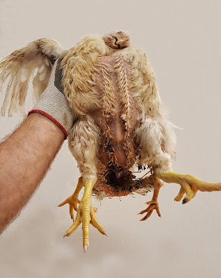
The state of polymelia (polymelus is the adjective) should not be confused with polydactilia. The former refers to an excessive number of limbs, the latter to an excess of digits. Polydactilia in birds has been associated with a genetic defect while polymelia results from incomplete division of co-twins soon after fertilization; its cause is not clear. Examples of polymelia in calves are shown elsewhere in LORI

Figure 1. A 7 week old polymelus broiler with duplication of the hind legs and probably the pelvis too. Image size: 1600 x 2500 px

The 7 week old broiler shown in figure 1 had a complete set of duplicate legs, situated caudal to the normal legs. Previously, this would have been termed a polymelus monster. Contemporary sensibility suggests that the term monster should not be used in such cases. The sex of this bird was not determined. 

In some cases of polymelia, the excess limb or limbs are separated from the skeleton of the surviving individual. In other cases, they are joined to its skeleton. Radiographic studies were not performed to ascertain any degree of skeletal fusion.

Several famous cases of humans with polymelia have been reported, including that of Myrtle Corbin.  In her case the form of polymelia resulted from a condition known as dipygus  (< Gr. di & pyge meaning "two buttocks"). This broiler too, could be described as dipygus; the two extra legs appeared to have a separate pelvis from that of the dominant bird.

There are numerous published reports of polymelia in poultry. In one case report, the extra set of limbs was surgically removed. This leaves one to ponder on the possible culinary fate of others.


Selected references:

Abu-Seida, A,M. 2014. Amputation of polymelia in a layer chicken. Avian Diseases: 58: 330-332.

Ajayi, I.E. and Mailafia, S. 2011. Occurrence of polymelia in 9-week-old male broiler: anatomical and radiological aspects. J. Vet.Anat. 4:69-67

Amatya, B. (2015). First Amatya, B. 2014 Report of polymelia in shakini breed chicken from Nepal. J. Nat. History Museum 28:175-177.

OL, A and Oyagbemi, A.A.2013. First report of polymelia and a rudimentary wing in a Nigerian Nera black chicken. J.SAVA 27:84

PoultryDVM. 2019. Polymelia http://www.poultrydvm.com/condition/polymelia


Monday, March 20, 2017

Rabbit semen collection and use.

Rabbit semen collection and use.

Keywords: rabbit, semen, collection, vagina, artificial, lagomorph

Compared with Europe, Asia and South America, rabbit farming is underdeveloped in North America. Therefore, it is only superficially addressed in most of our animal science and veterinary curricula. Nevertheless, rabbits are important as livestock; farmed for meat and of course, angora fiber. They also have unique value for humane scientific study (See: Foote and Carney, 2000). 

On one occasion, guided by ignorance, the author built this surprisingly effective artificial vagina (AV) to collect rabbit semen. It was based on a bovine AV but had a liner constructed from the finger of a surgical glove. A small hole on the side, covered by another piece of the glove was created to admit warm water and air as required. As can be seen, the liner terminated in a small collection vessel, cut from the conical tip of  plastic test tube. 


Image size: 1151 x 1563 px

Using a receptive doe, the process of semen collection was simple. Semen collection can be further simplified by training the buck to mount the skin of a doe, covering the hand that holds the AV. Copulation is rapid, resembling most closely, that seen in ruminants.

In the majority (~70%) of semen samples, a small plug of clear mucus will be found in the AV as shown below. This is a product of the vesicular glands. In sequential samples collected on the same day, the presence of a mucous plug is less likely (~5%). It was once surmised that the plug formed by this gel was essential to prevent loss of semen and produce normal conception rates. However it is now clear that conception rates can be excellent in the absence of any gel plug. Indeed, extended semen contains virtually no gel and conception rates using A.I. can be excellent. Therefore the presence of gel in rabbit ejaculates remains an enigma, rather like the gel in a stallion's ejaculate.


Image size: 1625 x 1099 px

To the author's eye, rabbit semen bears a striking resemblance to that of a bull; its motility and morphology being similar. Concentration is lower that of a bull's ejaculate however (~500 million per ml vs. 1000 million per ml). Nevertheless, wave motion (a function of both motility and concentration) can often be seen in rabbit ejaculates. 

Puberty in males is reached at six to seven months of age but peak sperm production may not be reached until 2 years of age or more.  At maturity, the volume of an ejaculate varies from 0.5 to 2 ml (occasionally more) but there is some variation at both upper and lower limits, partly due to significant volumes remaining on the liner of the AV.  For that reason, the AV is sometimes flushed after collection to retrieve more of the ejaculate.

Although sperm production increases under the effect of increasing day length in wild lagamorphs, this effect is not generally noticeable in domestic rabbits.

Although AVs are commercially available, readers may be interested to read about an alternative AV (Bredderman, 1964) that can be built for small scale breeding. Semen extenders and insemination pipettes are widely available for commercial rabbit production. Sources are readily available on the Internet.

Handling and insemination

Although acceptable fertility has been reported with doses as low as 0.5 million sperm, common practice dictates that between 20 and 40 million sperm are used per insemination, aiming for no more than 0.5 ml in volume to reduce sperm loss after insemination. A single ejaculate therefore, may provide between 20 and 50 insemination doses. 

For commercial use, semen from several males is pooled before insemination. The insemination pipette is bent dorsally to accommodate the ventrally-directed vagina and semen is simply deposited in the cranial vagina. No attempt is made to inseminate intra-cervically in either of the two cervixes. 

Frozen-thawed semen is seldom used in commercial rabbit production. Instead, ejaculates are diluted in commercial diluents, cooled to between 18 and 20 deg.C and used over a 24 to 48 hour period. 

To ensure that ovulation occurs (bearing in mind that it is predominantly mounting and not vaginal stimulation that induces ovulation in rabbits; Cf. cats) it is common to inject small intramuscular doses of GnRH analogs into the doe at the time of insemination. Because GnRH and its analogs are small molecules and readily absorbed through mucous membranes, some success has been achieved by adding GnRH analogs (usually buserelin) to insemination doses to induce ovulation. Without GnRH stimulation, the number of kits per litter is not commercially viable.

Selected references:

Alvarino, J.M.R. 2000. Reproductive performance of male rabbits World Rabbit Science Association, Volume 8, supplement 1. pp.13-35

Amann, R.P. 1966. Effect of ejaculation frequency and breed on semen characteristics and sperm output of rabbits. J. Reprod Fert. 11:291-293

Bredderman, P.J. 1964. An improved artificial vagina for collecting rabbit semen. J. Reprod. Fert. 7:401-403.

Brun, J.M. et al 2002. The relationship between rabbit semen characteristics and reproductive performance after artificial insemination. Anim. Reprod. Sci 70:139-149

Foote, R.H. and Carney E.W. 2000.The rabbit as a model for reproductive and developmental toxicity studies 

International rabbit reproduction group. 2005.Guideline for the handling of rabbit bucks and semen. World rabbit.Sci. 13:71-91

Lavara, R. et al. 2005. Do parameters of seminal quality correlate with the results of on-farm inseminations in rabbits? Theriogenology 64:1130–1141

Mocéa, E. and Vicenteb, J, 2009. Rabbit sperm cryopreservation: A review. Animal Reprod Sci. 110: 1–24

Morell, J.M. 1995. Artificial insemination in rabbits. British Vet. J. 151: 477-487

Mukherjee, D.P. et al 1951.The Gelatinous Mass in Rabbit Semen  Nature 168: 422 - 423

Theau-Clément, M. 2016. Relationships between rabbit semen characteristics and fertilising ability after insemination. Animal. 10:426-431

Vincente, J.S. et al. 2008. Rabbit reproductive performance after insemination with buserelin acetate extender. Livestock Science 115: 153–157 

Viudes-de-Castro, M.P.Ovulation induced by mucosa vaginal absorption of buserelin and triptorelin in rabbit 68: 1031-1036. Theriogenology 68: 1031–1036

http://placentation.ucsd.edu/rabbitfs.htm Kurt Benirschke.

Sunday, March 13, 2016

Zebra mare reproductive tract


Keywords: Equus, chapmani, Chapmans, Zebra, Equus quagga chapmani, uterus, ovaries


Note: Both zebra and zebras are widely accepted words as plurals for the singular form "zebra". The author has chose to use the term zebras for the plural form in this entry.

Although some disagreement exists on the taxonomy of Zebras there appear to be seven extant species of Zebras and a few subspecies within some of those species. Equus grevyi is the largest zebra and has a diploid chromosome number of 46. Plains zebras such as the Equus quagga chapmani featured in this LORI entry, have 44 chromosomes while Mountain zebras (Equus zebra zebra) have 32. Some zebras have finer striping than others, some differ in dentition, conformation and so on.

The reproductive biology and anatomy of horses and zebras are remarkably similar. 

Below, see an image of the reproductive tract of a mature (exact age unknown) Equus quagga chapmani. The parity of the mare was also unknown but examination of the tract suggested that she had been pregnant at least once. In this image, the cervix appears to be shorter than that of a domestic mare. However, detailed examinations of the reproductive tracts of 310 Equus quagga showed an average length of 4.9 cm; similar to than of domestic mares.


Image size:1412 x 872px

The image below accentuates the remarkable similarity between this animal and domestic mares with regard to the vestibulovaginal seal (arrows) i.e. the remnants of a hymen.


Image size:1064 x 654px

Again, below, the ovaries and adnexa of a Chapman's zebra are remarkably similar to those structures in domestic mares in both size and architecture. The author's fingertips have been inserted into the ovarian bursa.


Image size: 1384 x 1236px

This mare was examined in early January (in the northern hemisphere) when most domestic mares would have been in winter anestrus. This probably explains the lack of activity in her ovaries. Multiple ovulations are rare in zebras (twins being virtually unknown) but secondary ovulations do occur during pregnancy and accessory corpora lutea form as is the case in domestic mares.

Image size:2816 x 2112px

The author was struck by the similarity of the clitoris in this zebra and those in domestic mares. There was also at least one well developed clitoral sinus within dorsal surface of the clitoris. This led the authhor to contemplate the potential for Taylorella equigenitals or or other Taylorella species to cause reproductive failure in zebras.

 Image size:2816 x 2112px


General commentary:

Like Equus caballus, Zebras are seasonal breeders, responding to increased day length. However, there appears to a remarkable effect of increased energy availability in modulating that response to day length.

From numerous observation by many authors, zebra mares appear to have standing estrous periods that are similar in duration to those in domestic mares. Their gestation is however, substantially longer. Although gestation as short as 336 days has been recorded, most zebras foal after gestations of approximately 370 days. Normal gestation can be as long as 425 days.

Progesterone, eCG and estrogen profiles are similar to those in mares and certainly, urine estrogens shed during urination in sand bedding can be used to diagnose pregnancy. Similarly, assays for eCG in urine may hold promise for pregnancy diagnosis. Indeed, the latter has shown to be the case in domestic mares.

Mammary development, foaling, "foal heat" and neonatal physiology in zebras are similar to the same features in domestic horses but weaning of zebra foals only occurs after 10 to 12 months.

Interestingly, zebra of all stripes (!) can interbreed with domestic horse and donkeys and produce normal, live offspring. In many cases, those hybrid offspring are fertile as well.


Selected references:

Allen, W.R. and Short, R.V. 1997. Interspecific and extraspecific pregnancies in
equids: Anything Goes. J.Heredity 8:384-392

Asa, C.S. et al. 2001. Patterns of excretion of fecal estradiol and progesterone and urinary chorionic gonadotropin in Grevy’s zebras (Equus grevyi): Ovulatory cycles and pregnancy
Zoo Biology. 20: 185–195

Smuts, G.L. 1976. Reproduction in the zebra mare Equus burchelli antiquorum from the Kruger National Park Koedoe.19: 89-132


Sunday, January 4, 2015


Twinning in avian species


Keywords: avian, twins, triplets, freemartinism


Image size: 1352 x 734px

Note: this is a serious entry, only the image is somewhat tongue/egg-in-cheek

I was amazed to crack open two large eggs and later, to have two sets of twin yolks sitting there on my toast. Being compulsively academic, but essentially mammalian in orientation, I began to wonder about the incidence of twinning, identical twins, mixed sex twins conjoined fetuses and even Freemartinism in birds.

Here is a summary of what I learned:

Twinning appears to be common in birds, with twins arising from eggs with double yolks, eggs with one yolk and two blastoderms, or eggs with one yolk and a single blastoderm. In the last two cases, one presumes that the chicks are identical twins. Indeed, both fraternal and identical twins do occur from single eggs in birds. In rare cases, triplets and even quadruplets may occur. In a remarkable photograph from a paper published in 1940, H.H. Newman shows triplets and conjoined chicken embryos in their early stages of development.

Twins have been reported in many species of birds and may be induced by stress. In support of this statement, the incidence of twins rises in laying hens exposed to hypothermia.

As expected, individual twins weigh less than singlets and twins may be of the same or mixed sex. Vascular anastmoses develop during gestation in twin chickens and in mixed sex chickens a freemartin-like condition has been reported in female chicks.

References:

Bassett, S.M. et al. 1999 Genetically identical avian twins. Journal of Zoology. 247: 475–478

Burke, W.H. and Sharp, P.J. 1989. Sex Differences in Body Weight of Chicken Embryos. Poultry Science. 68: 805-810.

Griffith, S.C. and Stewart, R. 1998. Genetic confirmation of non-identical embryonic twins in the House Sparrow Passer domesticus. Journal of avian biology 29:207-208

Lutz, H. Lutz-Ostertag, Y. 1975. Intersexuality of the Genital System and “Free-Martinism” in Birds. pp. 382-391 in Intersexuality in the Animal Kingdom. Springer Berlin Heidelberg. Print ISBN 978-3-642-66071-9

Newman, H.H. 1940. Twin and triplet chick embryos. Journal of Heredity. 31: 370-378

Pattee, O.H. et al. 1984 Twin embryos in a peregrine falcon egg. The Condor 86:352-353


Monday, July 28, 2014

Murine placenta.

Keywords: mouse, placenta, intimate, histology, human


Image size:1920 x 902 px

This histologic image of a murine placenta shows the intimate relationship between the fetal and maternal compartments in this species, more intimate than in any of the domestic species. It is fascinating because the fetal and maternal compartments are so easy to differentiate. That is because of the presence of nucleated red blood cells (RBCs) in the fetal compartment (green arrow). In general, most fetal RBCs have nuclei and most maternal RBCs (yellow arrow) do not. Also, fetal RBCs are significantly larger than the maternal RBCs .
The reader is reminded that not all fetal red blood cells are nucleated, and that the proportion of nucleated RBCs varies through gestation and from one species to another. In young murine fetuses for example, the site of hematopoesis in is the liver, not the spleen or bone marrow and RBCs from the liver do not have nuclei. By contrast, in equine young fetuses virtually all RBCs are nucleated, declining to the time of foaling, when nucleated RBCs are rare. Also, some adult domestic animals such as new world camelids and some breeds of dogs (Schauzers and Dachshunds) normally have low nucleated RBC counts.

Anatomy: In the case of the mouse, the vessels of the maternal and fetal placentas intertwine with one another in an labyrinth of entanglement; a 'spongiotrophoblast' In the human, fetal villi are bath in maternal blood. that lies in spaces between the villi. The comparative histological structures of these placentas has been well illustrated E. Maltepe et al.

Human and murine placentas are similar; intimate and single discoid in gross anatomy. This explains why the mouse is often used as a model to study human placental function. Although the placentas are similar, in human placentation, blood in intervillous spaces baths the chorion of the fetus directly. In the mouse, maternal endothelial cells, not blood itself, come to lie against the chorion. Contrary to older data, in neither case are fetal endothelial cells exposed to maternal blood. Those data suggested that human placenta were hemo-endothelial, a situation that does not exist. Currently, human and murine placentas are respectively classified as hemo-chorial and endotheliochorial. As found in many placentas, multinucleate syncytiotrophoblasts (literally fused cells with many nuclei that feed and grow) are present at the interface of the fetus and endometrium. In the murine placenta two of these multinuceate layers separate the endothelia of the fetal and maternal capilliaries from one another. Only a few multinucleated cells (white arrow) can be seen in this image.

The large ringed cell is a  "giant cell" of fetal  origin  These are common in the placentas of rabbits, squirrels, marmots, rats and mice. They are highly invasive and interestingly,.usually polyploid, having up to four time the diploid number of  chromosomes in the rest of the  fetus! These are also important secretory cells, producing placental lactogen, growth hormone and cytokines that control placental re-modeling as the fetus grows.

The yellow arrow points to cells that are probably part of the so called junctional zone that lies between the labrinthe (closest to the fetus) and the dicidual zone, (Georgiades et al.) which is embedded in the endometrium.


References:

- Maltepe1, E. et al. 2010. The placenta: transcriptional, epigenetic, and physiological integration during development. J Clin Invest.120:1016–1025.
- Blood and Blood Formation Russell, E.S. and Bernstein S.E. http://www.informatics.jax.org/greenbook/frames/frame17.shtml
- Simmons G et al. 2007. Diverse subtypes and developmental origins of trophoblast giant cells in the mouse placenta. Developmental Biol.304:567–578
- Mossman, Harland, W. Vertebrate Fetal Membranes. ISBN: 0-8135-1132-1.
- Nuceated red blood cells: https://ahdc.vet.cornell.edu/sects/clinpath/modules/hemogram/nrbc.htm
- Allen A.L. et al. 1998 Hematology of Equine Fetuses with Comparisons to Their Dams Vet. Clin. Path. 27: 93-98.
Georgiades, P. aet al. 2001, Roles for genomic imprinting and the zygotic genome in placental development. Proc. Nat. Acad. Sci. 98: 4522–4527